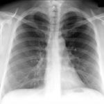
Radiography involves exposing a part of the body to a small dose of radiation to produce an image of the internal organs. When x-rays penetrate the body they are absorbed in varying amounts by different tissues. Ribs, for example, are dense and will block much of the radiation and, therefore, appear white or light gray on the image. Soft tissue such as the liver or lungs will appear darker because more radiation can pass through it to expose the film.
X-ray imaging is the fastest and easiest way for a physician to view and assess broken bones, joint or spine injuries. At least two images (from different angles) are taken and often three images are needed if the problem is around a joint (knee, elbow or wrist). X-rays also play a key role in guiding orthopedic surgery and in the treatment of sports-related injuries. X-ray may uncover more advanced forms of cancer in bones although early screening for cancer findings requires other methods.
Images are stored electronically using state-of-the-art PACS (Picture ArChiving System). Images may be digitally acquired or may be digitized from analog images to be stored on PACS.