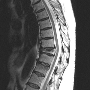
Every day, physicians use radiography, or x-rays, to view and evaluate bone fractures and other injuries of the musculoskeletal system. However, a plain x-ray test is not the best way to assess bone density. To detect osteoporosis accurately, doctors use an enhanced form of x-ray technology called dual-energy x-ray absorptiometry (DEXA). DEXA bone densitometry is today’s established standard for measuring bone mineral density (BMD). It is a quick, painless procedure for measuring bone loss. Measurements of the lower spine and hips are most often done.
The DEXA machine sends a thin, invisible beam of low-dose x-rays with two distinct energy peaks through your bones. One peak is absorbed mainly by soft tissue and the other by bone. The soft tissue amount can be subtracted from the total and what remains is a patient’s bone mineral density.
The amount of radiation used is extremely small—less than one-tenth the dose of a standard chest x-ray.
More portable devices that measure the wrist, fingers or heel are sometimes used for screening, including some that use ultrasound waves rather than x-rays.
Osteoporosis involves a gradual loss of calcium, causing the bones to become thinner, more fragile and more likely to break. The DEXA test can assess your risk for developing fractures. If your bone density is found to be low, you and your physician can work together on a treatment plan to help prevent fractures before they occur. DEXA is also effective in tracking the effects of treatment for osteoporosis or for other conditions that cause bone loss. Bone density testing is strongly recommended if you:
- are a post-menopausal woman and not taking estrogen.
- have a personal or maternal history of hip fracture or smoking.
- are a post-menopausal woman who is tall (over 5 feet 7 inches) or thin (less than 125 pounds).
- are a man with clinical conditions associated with bone loss.
- use medications that are known to cause bone loss, including corticosteroids such as Prednisone, various anti-seizure medications such as Dilantin and certain barbiturates, or high-dose thyroid replacement drugs.
- have type 1 (formerly called juvenile or insulin-dependent) diabetes, liver disease, kidney disease or a family history of osteoporosis.
- have high bone turnover, which shows up in the form of excessive collagen in urine samples.
- have a thyroid condition, such as hyperthyroidism.
- have experienced a fracture after only mild trauma.
- have had x-ray evidence of vertebral fracture or other signs of osteoporosis.
